
Understanding the EEG results for tonic‑clonic seizures can feel like decoding a foreign language, but with the right roadmap you’ll know what each spike, wave, and pause really means.
- Identify the three EEG phases that surround a generalized tonic‑clonic seizure.
- Learn the hallmark ictal pattern-high‑frequency polyspikes followed by a burst‑suppression.
- Distinguish true epileptiform activity from common artifacts.
- Apply ACNS‑recommended reporting standards.
- Use the EEG findings to guide treatment decisions.
What the test actually records
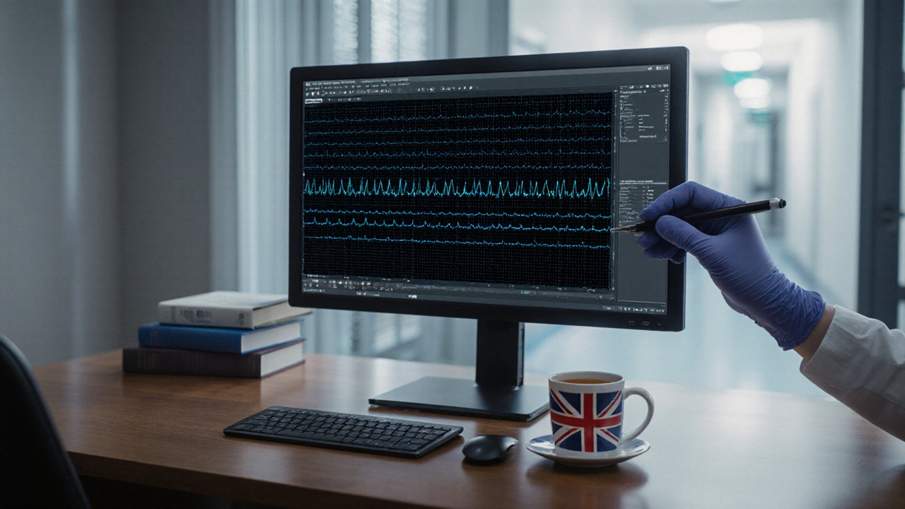
Electroencephalogram (EEG) is a non‑invasive neurophysiological test that records electrical activity of the brain via scalp electrodes, typically sampled at 500-1000Hz and displayed as voltage‑time traces. The system captures the summed postsynaptic potentials of cortical pyramidal cells, which means the signal reflects large‑scale neuronal synchrony rather than single‑neuron spikes.
Why tonic‑clonic seizures look the way they do on EEG
Tonic‑clonic seizure is a generalized epileptic event characterized by a tonic phase of muscle stiffening followed by a clonic phase of rhythmic jerking. During the tonic phase, cortical neurons enter a high‑frequency, low‑amplitude firing mode that translates on EEG to a burst of polyspikes-often 20-30Hz-lasting 0.5-2seconds. The subsequent clonic phase produces a rhythmic spike‑and‑wave pattern around 3Hz that mirrors the visible jerks.
Ictal EEG patterns - the “signature” of a tonic‑clonic event
The EEG interpretation of a generalized tonic‑clonic seizure hinges on three hallmarks:
- Polyspike burst: rapid series of spikes (often >10spikes/sec) that signal the onset of the tonic phase.
- Generalized spike‑and‑slow wave: 3‑4Hz spikes followed by a slow wave, coinciding with the clonic movements.
- Post‑ictal suppression: a period of low amplitude (<10µV) activity lasting 10‑30seconds as the brain recovers.
These features are distinct from focal seizures, which usually start with a localized rhythmic discharge that spreads gradually.
Polyspike burst is a rapid series of high‑frequency spikes that marks the onset of the tonic phase in a generalized seizure.
Interictal epileptiform discharges - what you see when the seizure isn’t happening
Interictal epileptiform discharge (IED) is a brief (<100ms) spike or sharp wave that occurs between seizures and signals an increased likelihood of future events. In patients with generalized tonic‑clonic seizures, IEDs typically appear as generalized 2-3Hz spike‑and‑wave bursts, even during wakefulness. Their presence supports a diagnosis of generalized epilepsy and can guide medication choice.
Post‑ictal EEG - the brain’s wind‑down period
Post‑ictal EEG is a recording captured after a seizure, showing a suppression of background activity followed by a gradual return to baseline. The depth and duration of suppression correlate with seizure severity; longer suppression often predicts a transient cognitive “recovery” phase. Recognizing this pattern prevents mislabeling the low‑voltage segment as a technical failure.
Guidelines that keep reports consistent
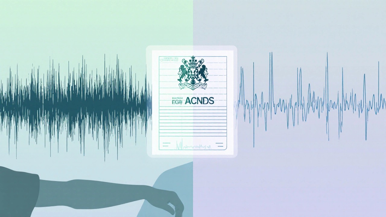
American Clinical Neurophysiology Society (ACNS) guideline is a consensus document that standardizes terminology, electrode placement, and reporting criteria for EEG in epilepsy. Since 2020, the guideline recommends explicitly noting the three phases (ictal, interictal, post‑ictal) and using the term “generalized polyspike burst” rather than vague descriptors. Following ACNS standards improves inter‑rater reliability and helps neurologists compare studies across centers.
Where the seizure starts - the concept of a seizure onset zone
Seizure onset zone is a brain region that first generates the abnormal electrical discharge during a seizure. In true generalized tonic‑clonic seizures, the onset zone is essentially the whole cortex, which is why the EEG shows simultaneous bilateral activity. Differentiating this from a rapidly propagating focal seizure can affect whether a patient receives broad‑spectrum antiepileptic drugs (AEDs) or considers surgical evaluation.
Common pitfalls - artifacts that mimic seizures
Artifact is a non‑physiological signal, such as muscle movement, eye blink, or electrode malfunction, that contaminates the EEG recording. Muscle (EMG) bursts during a tonic phase can look like high‑frequency polyspikes, but they usually have a broader spectral range and are localized near temporal electrodes. Eye‑blink artifacts generate slow waveforms that can be mistaken for post‑ictal suppression if the montage is limited. Knowing these red flags saves time and avoids unnecessary medication adjustments.
Putting it together - how clinicians act on the EEG
When a neurologist reads an EEG that shows the classic polyspike‑burst → generalized spike‑and‑wave → post‑ictal suppression sequence, they can confidently label the event as a generalized tonic‑clonic seizure. The report then informs several downstream decisions:
- Medication selection: drugs that enhance generalized inhibition (e.g., valproate, levetiracetam) are preferred.
- Risk stratification: frequent IEDs or prolonged post‑ictal suppression may signal higher seizure burden, prompting a dose increase.
- Driving clearance: a documented generalized seizure mandates a minimum three‑month seizure‑free period in most jurisdictions.
- Family counseling: visualizing the EEG pattern helps explain the condition to patients and caregivers.
Related concepts you’ll encounter next
Understanding EEG results opens doors to deeper topics such as sleep‑deprived EEG, which can unmask interictal spikes; quantitative EEG (qEEG), a computer‑based analysis that measures power spectra; and magnetoencephalography (MEG), an imaging technique that pinpoints the seizure onset zone with millimeter precision. Each builds on the basics covered here.
| Pattern | Typical Frequency | Duration | Clinical Correlate |
|---|---|---|---|
| Polyspike burst (ictal) | 20-30Hz | 0.5-2s | Tonic phase |
| Generalized spike‑and‑wave (ictal) | 3-4Hz | 2-10s | Clonic phase |
| Interictal epileptiform discharge | 2-3Hz (spike‑and‑wave) | <0.1s | Between seizures |
| Post‑ictal suppression | <1Hz (low amplitude) | 10-30s | Recovery period |
Frequently Asked Questions
Can a single EEG capture a tonic‑clonic seizure?
Yes, but the chance is low because generalized seizures are brief and often occur unpredictably. Long‑term video‑EEG monitoring or a sleep‑deprived study increases the odds of catching an event.
What does “burst‑suppression” mean on a post‑ictal EEG?
Burst‑suppression refers to alternating periods of high‑amplitude activity (bursts) and flat, low‑voltage background (suppression). In the post‑ictal state it reflects neuronal exhaustion after a massive synchronous discharge.
How can I tell muscle artifact from true polyspikes?
Muscle artifact usually shows a broader frequency range (up to 100Hz), appears most prominently in temporal or frontal leads, and often coincides with visible movement. Polyspikes are more uniform, confined to 20-30Hz, and are present across all channels simultaneously.
Why are interictal spikes important if the patient isn’t seizing?
Interictal spikes indicate an underlying hyper‑excitable cortex and predict future seizure risk. Their presence can justify initiating or adjusting antiepileptic medication even in the absence of recent clinical events.
Do antiepileptic drugs change the EEG pattern?
Effective AEDs often reduce the frequency and amplitude of interictal spikes and may shorten the post‑ictal suppression phase. However, some drugs (e.g., benzodiazepines) can introduce diffuse slowing, which must be distinguished from pathology.
Sadie Viner
September 27, 2025 AT 17:40When interpreting a generalized tonic‑clonic seizure, it helps to segment the record into three distinct phases: the pre‑ictal baseline, the ictal polyspike‑burst followed by the spike‑and‑wave, and the post‑ictal suppression. The polyspike burst typically occupies the initial half‑second to two seconds and signals the tonic onset. The subsequent 3‑4 Hz spike‑and‑slow wave mirrors the clonic activity, and finally the brain enters a low‑amplitude suppression that can last several seconds.
Recognizing this sequence improves both diagnosis and treatment planning.
Kristen Moss
September 29, 2025 AT 21:54That polyspike burst description is spot on.
Rachael Tanner
October 2, 2025 AT 02:08The high‑frequency polyspike burst acts like a neural fireworks display, heralding the sudden shift into the tonic phase. Following that, the rhythmic spike‑and‑wave pattern locks onto the motor jerks we observe clinically. Finally, the post‑ictal silence isn’t empty-it reflects cortical exhaustion and can guide antiepileptic dosing decisions.
Christopher Xompero
October 4, 2025 AT 06:22Yo, the EEGg shows a *burst‑supression* like a lightning strike, then a slow wave that’s almost a nap for the brain.
It’s kinda like the brain’s doing a quick sprint and then a slow walk.
Debra Laurence-Perras
October 6, 2025 AT 10:36Seeing the three phases laid out like this makes the EEG much less intimidating for newcomers. It also provides a clear checklist for anyone writing a formal report.
dAISY foto
October 8, 2025 AT 14:50OMG, the polyspike burst is literally the brain’s way of screaming “GET READY!” before the clonic dance begins.
Then the spike‑and‑wave rolls in like a drumbeat, matching each jerk you see on the patient.
After the party, the post‑ictal suppression is the brain’s chill‑out zone, a quiet after‑glow.
Remember, catching all three windows can totally change how you tweak meds.
Ian Howard
October 10, 2025 AT 19:04Understanding the electrophysiological signatures of a generalized tonic‑clonic seizure begins with appreciating the spatial and temporal resolution of modern EEG systems. Contemporary setups sample at 500‑1000 Hz, which ensures that the fast polyspike bursts-often exceeding 20 Hz-are captured without aliasing. The polyspike burst itself is generated by a sudden, synchronized depolarization of large cortical pyramidal populations, and its duration of 0.5‑2 seconds reflects the transition from a resting to a hyper‑excitable state. During this burst, the amplitude may climb to 200‑300 µV, but the waveform morphology remains distinct from muscle artefacts because of its consistent inter‑spike interval and scalp distribution. Immediately after the burst, the brain settles into a 3‑4 Hz spike‑and‑slow wave complex that mirrors the rhythmic clonic jerks observed clinically; each spike is followed by a slow wave of roughly 200‑300 ms, creating a “saw‑tooth” appearance on the trace. The spike component is generated by thalamocortical resonance, while the slow wave reflects cortical inhibition and the refractory period of neuronal membranes. It is crucial to differentiate this genuine ictal pattern from common artefacts such as electrode movement, eye blinks, or ECG interference; artefacts typically lack the bilateral, synchronous morphology that characterises true epileptiform activity. Post‑ictally, the EEG enters a suppression phase where amplitudes drop below 20 µV, indicating widespread neuronal silencing and metabolic recovery. The length of this suppression-anywhere from a few seconds to over a minute-has prognostic value, as prolonged suppression often correlates with higher seizure severity and may influence medication adjustments. According to the ACNS guidelines, each of these phases should be annotated separately, with precise timestamps and descriptive tags, to facilitate inter‑rater reliability and downstream analytics. When reporting, the clinician should note the onset latency of the polyspike burst relative to the clinical observation, the frequency and morphology of the spike‑and‑wave complex, and the duration of post‑ictal suppression. This structured approach not only satisfies reporting standards but also aids in tailoring antiepileptic therapy; for example, a prominent polyspike burst may suggest a need for drugs that target fast sodium channels. Moreover, longitudinal EEG monitoring can reveal whether these patterns evolve over time, which is valuable for assessing disease progression or treatment response. In practice, integrating these EEG insights with the patient’s clinical picture-such as seizure frequency, triggers, and comorbidities-creates a comprehensive management plan that optimizes outcomes.
Bottom line: meticulous phase identification, artefact discrimination, and adherence to ACNS reporting standards turn raw EEG data into actionable clinical intelligence.
Jason Lancer
October 12, 2025 AT 23:18The breakdown you gave really demystifies the spike‑and‑wave, especially the part about thalamocortical resonance.
Tony Bayard
October 15, 2025 AT 03:33Exactly, and when you pair that with the post‑ictal suppression duration, you can actually predict which drug will curb the next episode.
Irene Harty
October 17, 2025 AT 07:47The ACNS consensus underscores the necessity of annotating each ictal phase with unequivocal timestamps. Such rigor ensures that inter‑institutional studies maintain methodological fidelity.
Nymia Jones
October 19, 2025 AT 12:01It is incumbent upon every neurologist to adhere strictly to these annotation protocols; deviation compromises both diagnostic accuracy and patient safety.
Kiersten Denton
October 21, 2025 AT 16:15Seeing the phases laid out like a flowchart helps me stay focused during review.
Karl Norton
October 23, 2025 AT 20:29While the three‑phase model is useful, it sometimes glosses over subtle variations that could signal atypical seizure propagation. Clinicians should remain vigilant for those nuances.
Ashley Leonard
October 26, 2025 AT 00:43I love how the post highlights practical steps for distinguishing artefacts from true spikes. It makes the whole EEG interpretation feel more approachable.
Ramanathan Valliyappa
October 28, 2025 AT 03:57Artefact discrimination is essential for accurate diagnosis.
lucy kindseth
October 30, 2025 AT 08:11Just remember to double‑check electrode impedance before you start; low‑quality leads can mimic polyspikes and throw you off.
Karen McCormack
November 1, 2025 AT 12:25In the grand theatre of the brain, the polyspike burst is the opening act, a fleeting crescendo that announces the drama to come.
The spike‑and‑wave follows as the chorus, echoing the rhythmic pulse of existence.
Finally, the post‑ictal silence is the curtain call, a moment of reflection before the next performance.
Earl Hutchins
November 3, 2025 AT 16:40Mark each phase clearly. Follow ACNS rules.